

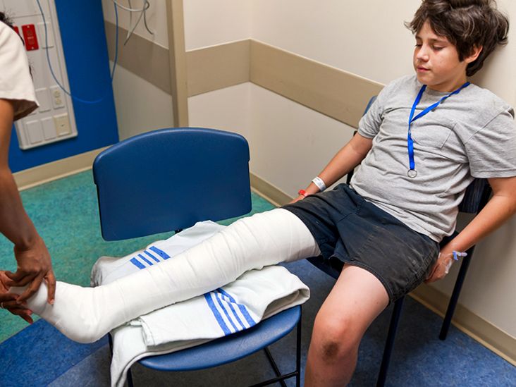
Fat embolism sounds like something that happens after eating too many fries, but thankfully, your lunch is not the villain here. A fat embolism occurs when tiny droplets of fat enter the bloodstream and travel to small blood vessels, most often in the lungs. In many cases, these droplets cause no noticeable trouble. But when they trigger breathing problems, brain-related symptoms, skin changes, and a body-wide inflammatory reaction, the condition is called fat embolism syndrome, or FES.
Fat embolism syndrome is uncommon, but it matters because it can become serious quickly. It is most often linked to trauma, especially fractures of long bones such as the femur, tibia, or pelvis. It may also occur after orthopedic surgery, severe burns, pancreatitis, sickle cell crisis, bone marrow procedures, liposuction, or other situations where fat or bone marrow particles enter circulation. The condition is tricky because its symptoms can look like other emergencies, including pulmonary embolism, pneumonia, sepsis, stroke, or acute respiratory distress syndrome. In other words, FES has a talent for wearing medical disguises.
This guide explains what causes fat embolism, how symptoms appear, who is most at risk, how doctors diagnose and treat it, and what can be done to reduce the risk. The goal is not to turn you into a trauma surgeon before breakfast, but to help you understand the essentials in plain, useful language.
What Is a Fat Embolism?
A fat embolism is a blockage or irritation in the blood vessels caused by fat droplets. These droplets usually come from bone marrow after a fracture, although other sources are possible. The fat can travel through veins to the lungs, where it may interfere with oxygen exchange. In some cases, droplets may pass through the lung circulation or a heart opening and reach the brain, skin, kidneys, retina, or other organs.
It is important to separate two related ideas:
Fat Embolism vs. Fat Embolism Syndrome
Fat embolism means fat droplets are present in the bloodstream. This can happen after trauma and may cause no obvious symptoms. Fat embolism syndrome means those fat droplets have caused a recognizable illness, usually involving the lungs, brain, and skin. Think of fat embolism as the “event,” and fat embolism syndrome as the “problem that may follow.”
Fat embolism syndrome usually appears within 24 to 72 hours after injury, especially after long-bone or pelvic fractures. However, timing can vary. A patient may seem stable at first, then develop shortness of breath, confusion, fever, rapid heartbeat, or a dotted rash a day or two later. That delayed appearance is one reason doctors watch trauma patients closely even after the broken bone has been identified and stabilized.
What Causes Fat Embolism?
The most common cause of fat embolism syndrome is orthopedic trauma. Long bones contain fatty marrow. When a bone breaks, pressure inside the bone can force marrow fat into damaged blood vessels. Those fat droplets then move through circulation, like tiny uninvited guests looking for the wrong party.
Common Causes
The leading causes and triggers include:
- Femur fractures: The thigh bone is one of the classic injuries associated with FES.
- Tibia fractures: Lower-leg fractures can also release marrow fat.
- Pelvic fractures: These injuries may involve significant trauma and bleeding risk.
- Multiple long-bone fractures: More injured marrow can mean more opportunity for fat droplets to enter circulation.
- Orthopedic procedures: Surgery involving the marrow cavity, such as intramedullary nailing or joint replacement, can increase intramedullary pressure.
- Severe soft-tissue injury or burns: Major trauma can trigger inflammation and fat release from tissues.
- Liposuction or fat grafting: Rarely, cosmetic procedures involving fat manipulation may lead to embolic complications.
- Bone marrow biopsy or transplant: These procedures involve marrow tissue and can rarely be associated with fat embolism.
- Sickle cell crisis: Bone marrow injury or necrosis may release fat into circulation.
- Acute pancreatitis: Fat metabolism and inflammation may contribute to embolic risk in rare cases.
How Fat Droplets Cause Damage
Experts generally describe two overlapping processes. The mechanical theory says fat droplets physically block tiny blood vessels, especially in the lungs. The biochemical theory says fat breaks down into free fatty acids that irritate blood vessels, damage the lung lining, and trigger inflammation. In real life, the body rarely reads one textbook chapter at a time, so both mechanisms may happen together.
The lungs are commonly affected because blood from the veins passes through the pulmonary circulation. When the lung’s small vessels become blocked or inflamed, oxygen levels can fall. In severe cases, this can progress to acute respiratory distress syndrome, a dangerous form of lung failure requiring intensive care.
Who Is Most at Risk?
Fat embolism syndrome is uncommon, but risk rises in certain situations. People at higher risk include patients with:
- Closed long-bone fractures, especially femur fractures
- Multiple fractures
- Pelvic fractures
- Delayed fracture stabilization
- Severe trauma involving bone and soft tissue
- Orthopedic surgery involving the marrow cavity
- Young adult age in some trauma patterns, often because high-energy injuries are more common in this group
- Underlying conditions that affect bone marrow or circulation, such as sickle cell disease
Not every person with a femur fracture develops FES. In fact, most do not. But because the syndrome can be dangerous, trauma teams pay attention to early warning signs. Prevention often begins with good fracture care, timely stabilization, oxygen monitoring, and careful supportive management.
Symptoms of Fat Embolism Syndrome
The classic pattern of fat embolism syndrome includes three major categories: respiratory symptoms, neurological symptoms, and a petechial rash. Not everyone has all three. Some patients have only subtle signs at first, which is deeply inconvenient because medicine already has enough mystery novels.
Breathing Symptoms
Respiratory problems are often the most important feature. Symptoms may include:
- Shortness of breath
- Rapid breathing
- Low oxygen levels
- Chest discomfort
- Cough
- Blue or gray coloring around the lips or fingers in severe cases
- Need for oxygen support or mechanical ventilation
Because these symptoms can resemble blood clot pulmonary embolism, pneumonia, or lung contusion after trauma, doctors use the full clinical picture rather than relying on one symptom.
Brain and Nervous System Symptoms
Neurological symptoms can appear when fat droplets or inflammatory changes affect the brain. These may include:
- Confusion
- Restlessness or agitation
- Drowsiness
- Headache
- Seizures
- Weakness or abnormal movements
- Coma in severe cases
These symptoms can be especially alarming after trauma because doctors must consider many possibilities, including head injury, low oxygen, shock, medications, infection, or stroke. Fat embolism syndrome is one possible explanation, particularly when neurological changes occur with low oxygen and a recent long-bone fracture.
Skin Symptoms: Petechial Rash
A petechial rash is a pattern of tiny red, purple, or brown pinpoint spots caused by small areas of bleeding under the skin. In fat embolism syndrome, it often appears on the upper chest, neck, shoulders, armpits, inside the mouth, or around the eyes. The rash may be brief and easy to miss, so it is not always present by the time someone checks.
Other Possible Signs
Additional signs may include fever, fast heartbeat, low platelet count, anemia, retinal changes, kidney issues, or abnormal lab results. These findings are not specific to FES, but they can support the diagnosis when combined with the right clinical setting.
When Is Fat Embolism an Emergency?
Fat embolism syndrome can be life-threatening. Seek emergency care immediately if someone develops breathing trouble, confusion, seizure, fainting, blue lips, severe drowsiness, or a new pinpoint rash after a fracture, orthopedic procedure, severe injury, or fat-related cosmetic procedure.
After trauma, it is tempting to focus only on the visible injury. A broken leg is obvious; low oxygen is quieter. That is why symptoms such as rapid breathing, sudden confusion, or unexplained fever after a fracture should never be brushed off as “just stress.” The body may be waving a small red flag, and in this case, the flag may come with petechiae.
How Doctors Diagnose Fat Embolism Syndrome
There is no single perfect test for fat embolism syndrome. Diagnosis is usually clinical, meaning doctors combine the patient’s injury history, symptoms, physical exam, oxygen levels, imaging, and lab results. This can feel frustrating, but it is common in complex trauma medicine. The body does not always provide neat labels like a grocery aisle.
Clinical Criteria
Several diagnostic criteria have been proposed, including Gurd and Wilson criteria, Schonfeld’s scoring system, and Lindeque’s criteria. These systems consider features such as respiratory distress, brain symptoms, petechial rash, fever, fast heart rate, low platelets, anemia, and other changes. They are helpful, but doctors still rely heavily on judgment.
Tests That May Be Used
Possible tests include:
- Pulse oximetry: Measures oxygen saturation.
- Arterial blood gas: Checks oxygen, carbon dioxide, and acid-base balance.
- Chest X-ray: May show lung changes, though early results can be normal.
- CT scan: Helps rule out other conditions such as blood clot pulmonary embolism or chest trauma.
- MRI brain: May show patterns consistent with cerebral fat embolism in patients with neurological symptoms.
- Blood tests: May reveal anemia, low platelets, inflammation, or organ stress.
- Eye exam: May detect retinal changes in some cases.
Because symptoms overlap with other conditions, diagnosis often includes ruling out more common or immediately treatable emergencies. A patient with low oxygen after a fracture may be evaluated for pulmonary embolism, pneumonia, aspiration, shock, lung contusion, medication effects, or sepsis at the same time.
Treatment for Fat Embolism Syndrome
There is no magic “fat embolism antidote.” Treatment is mainly supportive care, which means doctors support the body while the condition improves and prevent complications along the way. Supportive care may not sound glamorous, but in critical care, it is often the difference between danger and recovery.
Oxygen and Breathing Support
The first priority is oxygen. Mild cases may require supplemental oxygen through a nasal cannula or mask. More serious cases may require noninvasive ventilation or mechanical ventilation in an intensive care unit. If acute respiratory distress syndrome develops, doctors may use lung-protective ventilation strategies and careful fluid management.
Stabilizing the Injury
Proper fracture stabilization is a major part of management. Movement at the fracture site can continue to release marrow contents and worsen inflammation. Depending on the patient’s condition, stabilization may involve splinting, external fixation, or surgery. Trauma teams balance early fixation with overall stability, bleeding risk, and other injuries.
Fluids, Monitoring, and ICU Care
Patients may need intravenous fluids, blood pressure support, fever management, seizure treatment, and close monitoring of oxygen levels, blood counts, kidney function, and neurological status. Severe cases may require ICU care. Rarely, advanced support such as extracorporeal membrane oxygenation may be considered for extreme respiratory failure.
What About Steroids?
Corticosteroids have been studied for prevention and treatment, especially in high-risk orthopedic trauma patients. Some studies suggest they may reduce the risk of FES, but routine use remains debated because dosing, patient selection, and risk-benefit balance are not universally settled. In plain English: steroids are not a DIY prevention plan, and they are not automatically used for everyone with a broken bone.
Can Fat Embolism Be Prevented?
Prevention focuses on reducing the chance that fat droplets enter circulation and minimizing the inflammatory response after injury. Not every case can be prevented, but several strategies may lower risk.
Early Fracture Stabilization
Timely stabilization of long-bone and pelvic fractures is one of the most important preventive measures. Stabilization reduces movement at the fracture site and may reduce additional marrow fat release. In unstable trauma patients, temporary external fixation may be used first, with definitive surgery delayed until the patient is safer for a longer procedure.
Careful Surgical Technique
During orthopedic procedures, surgeons may use techniques that reduce pressure inside the bone canal and limit embolic load. The exact approach depends on the fracture type, patient condition, equipment, and surgical plan. This is one reason orthopedic trauma care is not a “watch a video and wing it” hobby.
Good Trauma and Critical Care
Prevention also includes oxygen monitoring, adequate hydration, blood pressure support, pain control, and early recognition of deterioration. In high-risk patients, nurses and physicians watch for subtle signs such as increasing breathing rate, falling oxygen saturation, confusion, fever, or rash.
Reducing Injury Risk in Daily Life
Many fat embolism cases are linked to accidents, so general injury prevention matters. Use seat belts, wear appropriate protective gear during sports or motorcycle riding, prevent falls at home, treat osteoporosis when present, and follow safety rules at work. No one can bubble-wrap life completely, but we can at least stop pretending ladders are furniture.
Recovery and Prognosis
Many people with fat embolism syndrome recover fully, especially with early recognition and good supportive care. Recovery time depends on severity. Mild cases may improve within days. Severe cases involving respiratory failure, seizures, or coma may require ICU care and a longer rehabilitation period.
Possible complications include acute respiratory distress syndrome, prolonged low oxygen, neurological injury, kidney stress, or, rarely, death. The outlook is generally better when the condition is recognized early, oxygen is supported promptly, and the underlying fracture or trigger is managed properly.
Living After Fat Embolism Syndrome
After recovery, follow-up may involve trauma surgery, orthopedics, pulmonology, neurology, rehabilitation medicine, or primary care. Patients may need physical therapy for the fracture, breathing exercises after lung injury, cognitive follow-up after neurological symptoms, or emotional support after a frightening ICU experience.
It is also normal for patients and families to have questions afterward. “Why did this happen?” “Could we have prevented it?” “Will it happen again?” The answers depend on the original injury, treatment timeline, and individual risk factors. A follow-up appointment is the best place to review the case in detail and get advice tailored to the person, not just the diagnosis.
Practical Experience: What Fat Embolism Looks Like in Real-Life Care
In real-world trauma care, fat embolism syndrome often begins as a puzzle rather than a dramatic announcement. Imagine a young adult brought to the emergency department after a motorcycle crash. The obvious problem is a femur fracture. The leg is painful, swollen, and clearly not applying for any dance competitions. Initial scans are done, pain medicine is given, and the orthopedic team plans stabilization. For the first several hours, the patient may seem alert and stable.
Then, perhaps the next day, small changes appear. The breathing rate climbs. Oxygen saturation dips. The patient seems unusually restless or confused. A nurse notices tiny red-purple spots near the shoulders or chest. None of these signs alone proves fat embolism syndrome, but together with a long-bone fracture, they raise concern. That is when the care team starts connecting the dots.
Families may feel blindsided because the broken bone looked like the main issue. This is completely understandable. A fracture is visible and concrete; fat droplets in tiny blood vessels are not exactly photogenic. But trauma affects the whole body. Bone marrow, inflammation, oxygen levels, blood pressure, and the lungs can all become part of the same story.
One practical lesson from fat embolism care is that monitoring matters. A patient after major fracture is not simply “waiting for surgery.” They are being watched for changes in breathing, mental status, circulation, pain, bleeding, and infection risk. When a nurse checks oxygen saturation again and again, it is not because the machine enjoys beeping. It is because early changes can guide fast action.
Another experience-based lesson is that communication helps. Families should tell the medical team if they notice new confusion, unusual sleepiness, trouble breathing, rash, or seizure-like activity. They should also ask simple, direct questions: “Is oxygen level okay?” “Could this be related to the fracture?” “What warning signs are you watching for?” Good questions do not annoy good clinicians; they help everyone stay focused.
For patients recovering from FES, the emotional side can be surprisingly strong. Some remember waking up in the ICU or being told they had a complication they had never heard of. Others remember very little and feel confused by the seriousness of what happened. Recovery may include gratitude, frustration, fear, and impatience all standing in line at once. That is normal. Healing from the fracture may take weeks or months, and healing from the scare may take time too.
Physical rehabilitation is often the most visible part of recovery. Depending on the injury, patients may need crutches, walkers, strengthening exercises, breathing exercises, or gradual return-to-work planning. The key is to follow the medical team’s restrictions. “I feel fine” is not the same as “my femur is ready for heroic nonsense.” Bones need time, lungs need patience, and the body appreciates not being rushed like a late airport passenger.
Prevention also becomes more personal after someone has seen fat embolism up close. Seat belts, fall prevention, safe driving, workplace protection, bone health, and prompt fracture care stop sounding like boring public-service wallpaper. They become practical tools. For older adults, preventing falls at home can be especially important: remove loose rugs, improve lighting, use handrails, check vision, review medications that cause dizziness, and treat osteoporosis when recommended.
For athletes and active people, protective equipment and smart training matter. For drivers and riders, helmets, seat belts, sober driving, and speed control matter. For anyone undergoing orthopedic surgery or cosmetic fat procedures, choosing qualified medical professionals and following pre- and post-procedure instructions matters. These steps do not guarantee perfect safety, but they lower riskand lowering risk is a very respectable life strategy.
The biggest takeaway from real-life experience is this: fat embolism syndrome is rare, but it rewards awareness. Patients do not need to diagnose themselves. Families do not need to memorize diagnostic criteria. But recognizing that breathing changes, confusion, seizure, or a pinpoint rash after major fracture can be serious may lead to faster care. In medicine, timing is often the quiet hero.
Conclusion
Fat embolism occurs when fat droplets enter the bloodstream, most often after long-bone or pelvic fractures. When those droplets trigger breathing problems, neurological changes, skin rash, and systemic inflammation, the condition is called fat embolism syndrome. Although uncommon, FES can be serious and may require oxygen support, intensive monitoring, fracture stabilization, and ICU care.
The most important points are straightforward: watch closely after major fractures, take new breathing or mental status changes seriously, stabilize fractures appropriately, and seek emergency care for warning signs. Fat embolism syndrome may be rare, but when it appears, fast recognition and supportive care can make a major difference.