There are ordinary repair jobs, there are ambitious hardware mods, and then there is whatever category applies when someone looks at a scanning electron microscope and thinks, “Yes, but what if this giant vacuum-powered dragon also talked nicely to a modern computer?” That, in a nutshell, is why Ben Krasnow’s SEM project grabbed so much attention. It was not just a geeky stunt. It was a clever act of translation between eras: one foot in the analog lab culture of knobs, phosphor screens, and slow scans, and the other in the digital maker world of microcontrollers, custom signal capture, and real-time processing.
Ben Krasnow has built a reputation for projects that feel part engineering, part detective novel, and part “please do not try this unless you really know what all these voltages are doing.” His work around electron microscopes fits that pattern perfectly. On one side, he documented a homebuilt scanning electron microscope project that proved he understood the machine at the level of beam formation, detection, noise control, and image generation. On the other, his later microscope hacking work showed a different kind of intelligence: not just building from scratch, but modernizing older lab equipment so it could produce better, more convenient digital results.
That is what makes the story behind [Ben Krasnow] Hacks A Scanning Electron Microscope so compelling. It is not merely about one instrument. It is about how curiosity, electronics know-how, and a refusal to accept “that’s just how old equipment works” can breathe new life into a machine that was never designed for the USB age. The result is a story that appeals to engineers, tinkerers, microscopists, and anyone who has ever looked at outdated hardware and muttered, “We can fix this.”
Why This Hack Turned Heads
Hackers love old hardware, but electron microscopes live in a different league from thrift-store cassette decks and rescued oscilloscopes. A scanning electron microscope, or SEM, is a serious scientific instrument. It uses a focused electron beam, a vacuum chamber, and multiple detectors to build magnified images from signals emitted by the sample. In plain English: instead of using light like a regular microscope, it throws electrons at tiny things and reads the fallout like a highly educated fortune teller.
That sounds dramatic because, frankly, it is. Even older SEMs are intricate systems. They need beam control, stable power, decent vacuum, careful signal collection, and image-generation hardware that all play nicely together. Krasnow’s hack drew attention because he did not treat the microscope like an untouchable relic. He treated it like a system that could be understood, probed, improved, and reinterpreted.
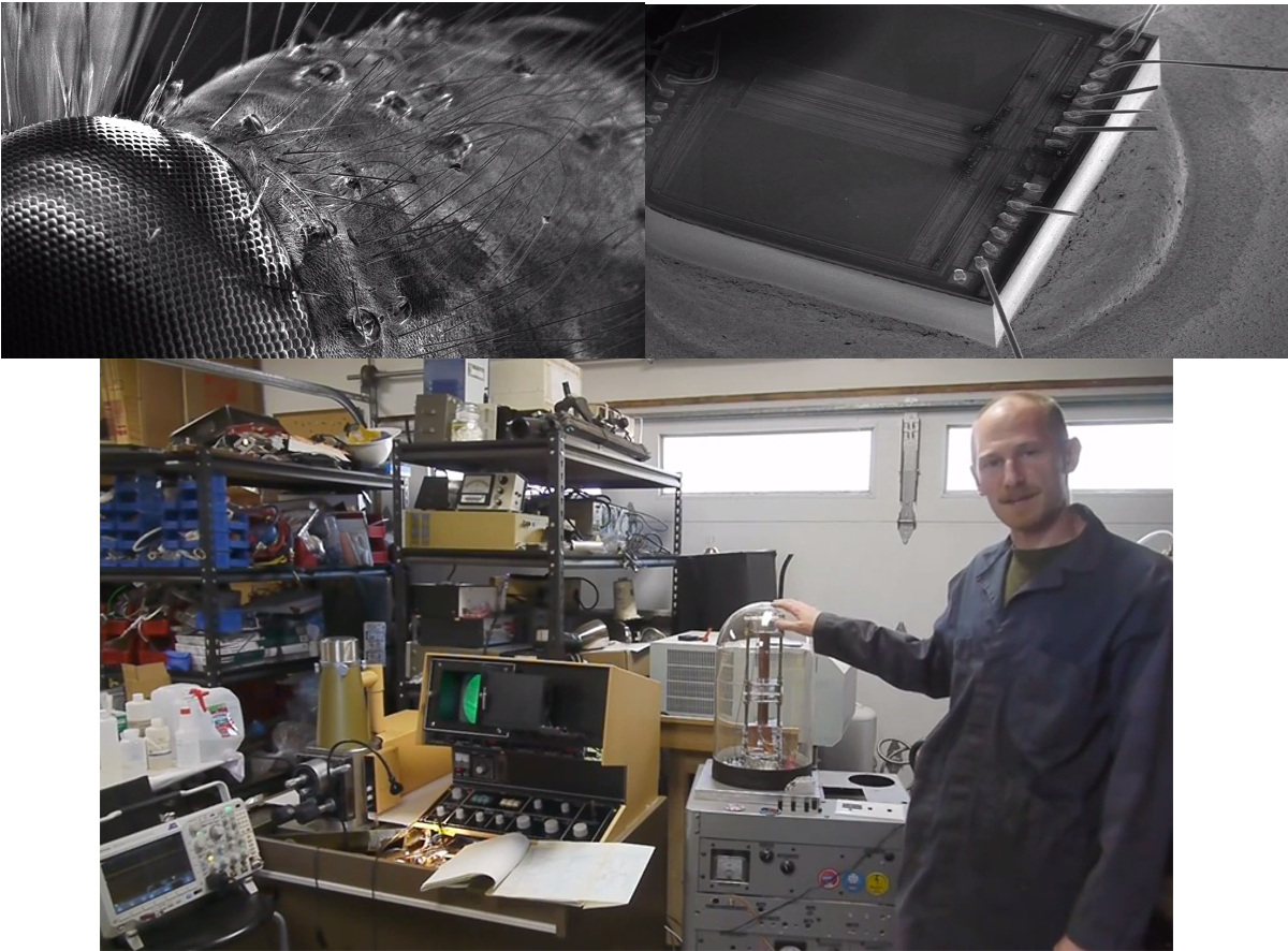
One of the memorable details from the coverage is that the older JEOL JSM-T200 could store images the old-fashioned way: through a screen-mounted Polaroid camera. That is charming in the same way rotary phones are charming, right up until you need to do actual work. In live mode, SEM images often look soft and ghostly because the instrument has very little time to collect signal at each point. Slow-scan photo modes improve image quality by spending more time gathering electrons, but they belong to an older workflow. Krasnow recognized that the instrument already had better image potential buried inside it. The trick was to capture that information digitally instead of settling for a legacy recording path.
What a Scanning Electron Microscope Actually Does
The beam, the vacuum, and the grayscale magic
To appreciate the hack, it helps to understand why SEMs work the way they do. An SEM forms an image by scanning a focused electron beam across a sample surface in a raster pattern. As the electrons interact with the sample, they generate different signals. Secondary electrons tend to carry topographic information from the near-surface region, while backscattered electrons can reveal compositional differences. Add X-ray signals and you can also investigate elemental makeup using EDS or EDX analysis.
This is why SEM images feel so different from ordinary photographs. They are not snapshots in the casual sense. They are constructed signal maps. Every bright ridge, dark trench, and suspiciously dramatic crater is the result of beam-sample interactions translated into brightness values on a screen. It is science wearing a grayscale trench coat.
Modern SEM references often stress that secondary electrons are especially useful for surface detail, while backscattered electrons are great for compositional contrast. That matters because it explains why microscope users obsess over detector choice, accelerating voltage, working distance, sample conductivity, and signal-to-noise ratio. They are not just fiddling with settings for fun. They are deciding what kind of truth the image will tell.
Why old SEM images can look eerie in live mode
The famous “ghostly” look of live SEM video is not a design flaw so much as a physics tax. In fast live scanning, the system has limited time to collect signal at each point. Fewer detected electrons generally means lower contrast and more noise. Slow scans fix that by allowing more signal accumulation, which produces cleaner, sharper images. That is why old photo modes could outperform the live display: the machine was not suddenly smarter, just more patient.
Krasnow’s insight was that patience plus digital capture could turn an old instrument into something much more practical. Instead of treating the microscope’s better imaging mode as a dead-end relic, he treated it as hidden bandwidth waiting to be liberated.
The Hack: Turning a Polaroid-Era Workflow Into a Digital One
This is where the project stops being merely “cool” and becomes genuinely instructive. Krasnow did not just bolt a camera onto a screen and call it innovation. He went inside the signal path. He identified usable video information in the microscope electronics, digitized it, handled sync, and sent the data to a computer for display and saving. In later documentation of his electron microscope capture system, he used a Teensy-LC-based setup to digitize analog output, extract video sync, and move the data over USB so software on the computer could reconstruct the image in real time.
That may sound straightforward if you say it quickly and avoid eye contact with the timing requirements. In reality, it is the sort of job that punishes sloppy assumptions. The signal must be sampled consistently. The timing has to be stable enough that the raster image does not turn into abstract art. Voltage levels often need translation before a microcontroller can safely read them. And all of that sits beside a machine that is already balancing beam control, vacuum, and detector behavior like a nervous circus act with electrons.
What makes Krasnow’s approach so satisfying is that it respects the microscope instead of replacing it. He did not build a fake SEM experience on top of the machine. He preserved the instrument’s underlying imaging logic and simply modernized the acquisition path. That is the difference between a gimmick and a real engineering upgrade.
Why the Project Resonates With Makers and Scientists
There is a broader lesson in this story, and it is bigger than one microscope. Scientific instruments often age unevenly. The core physics may still be excellent, while the user interface, storage method, or data workflow feels like it wandered in from a different century. A good hack identifies which parts of the system are still valuable and which parts are only hanging around because nobody has had the time, skill, or audacity to replace them.
That is exactly what happened here. The microscope’s imaging capability was not the problem. The bottleneck was how the image was being captured and used. By modernizing that layer, Krasnow effectively extended the practical life of the instrument and made it more useful for experiments, demonstrations, and creative imaging.
This also explains why his SEM work resonates so strongly with engineers. It celebrates a mindset that is equal parts reverse engineering and respect for the original design. He did not sneer at the old machine. He listened to it, figured out what it was trying to do, and then gave it a better way to communicate.
The Real Challenges Behind Any SEM Hack
Noise, stability, and the cruel honesty of electrons
Electron microscopes are brutally honest instruments. If your power supply ripples, your beam can wobble. If your scan linearity is off, shapes distort. If your detector path is noisy, the image turns mushy. Krasnow’s earlier SEM build notes show exactly this kind of iterative struggle: improving image quality by switching to a more linear X/Y deflection amplifier, tightening up shielding, and stabilizing power to eliminate rolling artifacts and focus problems.
That kind of troubleshooting is the real story behind almost every beautiful micrograph. The public sees the final image. The builder sees the list of sins that had to be confessed and corrected first.
Sample prep: where tiny things become drama queens
SEM imaging is also heavily influenced by the sample itself. Nonconductive materials can charge under the beam, creating bright artifacts and distorted information. Beam-sensitive materials can heat or degrade. That is why conductive coatings such as gold, platinum, or carbon are often used, and why low-kV imaging has become so important for delicate or insulating specimens. Lower accelerating voltages can reduce damage, improve surface sensitivity, and sometimes let users avoid heavy coating that would otherwise mask fine detail.
In other words, even if you hack the data path brilliantly, the electrons still get the final vote. SEM is a partnership between instrument design and sample behavior, and the sample is occasionally a diva.
More Than Pretty Pictures: What You Can Learn From the Images
Krasnow’s microscope work stands out because the images are not just decorative. They reveal structure in a way that makes the invisible feel mechanical and tangible. A window screen becomes a clean geometric test pattern for scan linearity. A MEMS gyroscope looks like a tiny engineered city. A drill bit becomes a sculpture of stress, wear, and texture. A vinyl groove transforms from familiar audio media into a microscopic landscape with enough detail to make you rethink how sound is stored.
That educational value matters. SEM images are powerful not only because they magnify, but because they reorganize intuition. They teach you that “smooth” surfaces are often chaotic, that tiny devices have architecture, and that manufacturing marks tell stories. Once you have seen an everyday object under an electron microscope, it becomes difficult to go back to trusting your unaided eyes like they are the final authority on anything.
This is one reason Krasnow’s microscope projects cross over so well into broader maker culture. They are technical, yes, but they are also visual explanations of the hidden physical world. The machine becomes both instrument and narrator.
Why This Story Still Feels Fresh
There is something timeless about combining old scientific hardware with modern embedded systems. It scratches several itches at once: preservation, problem-solving, performance improvement, and the sheer fun of proving that yesterday’s machinery still has tomorrow inside it. Krasnow’s SEM hack sits right in that sweet spot.
It also reminds us that innovation is not always about inventing a brand-new tool. Sometimes it is about noticing that a great tool has been trapped inside an outdated workflow. Free the workflow, and suddenly the machine feels new again.
That is the real charm of [Ben Krasnow] Hacks A Scanning Electron Microscope. It is a story about instrument hacking, but also about engineering optimism. The underlying message is wonderfully simple: if a machine still has something to teach, it is probably worth listening to before sending it off to become expensive furniture.
Experiences Around a Hacked SEM: What the Project Feels Like Up Close
If you spend time around a project like this, the experience is very different from the polished way it appears online. On a screen, a hacked scanning electron microscope looks like a clean success story: old machine, clever modification, sharp images, applause. In practice, it feels more like entering a conversation with a temperamental alien appliance that communicates through grayscale hints and occasional passive-aggressive noise.
The first emotional shift comes when you stop thinking of the SEM as one machine and start seeing it as a stack of subsystems. There is the vacuum system, which is already dramatic enough. There is the beam column, which behaves like a tiny ruler for the laws of physics. There is the detector and signal chain, where the microscope stops being pure hardware and starts becoming interpretation. Then there is the hacked acquisition path, where modern electronics try to make sense of signals born in an earlier design era. Every improvement feels like translating one dialect of engineering into another.
That is why the wins feel so satisfying. A cleaner signal is not just a cleaner signal. It is proof that the machine is becoming understandable. When a distorted image becomes straight, when rolling noise disappears, or when a faint ghostly pattern finally snaps into useful detail, it feels less like clicking a software setting and more like negotiating peace with physics. The microscope has not become easy. It has simply agreed, for a moment, to cooperate.
There is also something oddly theatrical about the objects you place inside. Ordinary items become performers. A mesh, a chip die, a worn drill bit, even a record groove suddenly carry the burden of revealing whether your setup is behaving properly. Under SEM imaging, familiar materials lose their social identity and become topography, composition, edge contrast, charging behavior, and signal response. A boring object can become thrilling if it tells you something honest about focus, detector geometry, or scan stability.
And then there is the psychological rhythm of the work. Projects like this reward stubborn patience. You make a change, test it, stare at a grayscale image, and ask yourself whether that brighter edge is real structure, a detector bias effect, a charging artifact, or your own wishful thinking wearing safety goggles. You learn to become suspicious in a productive way. That mindset is part of the pleasure. It turns every image into both result and question.
Most of all, a hacked SEM project changes how you look at tools. It becomes impossible to believe that equipment is fixed forever in the form it was sold. Once you have seen someone modernize image capture, rethink detector handling, or revive old microscope workflows with microcontrollers and software, you start seeing dormant potential everywhere. The experience is inspiring because it proves that expertise is not just about using advanced machines. Sometimes it is about refusing to let a brilliant machine stay trapped in yesterday’s habits.
That is why Ben Krasnow’s microscope work keeps circulating years later. It offers more than technical entertainment. It captures the feeling of standing at the border between analog craftsmanship and digital control, then deciding to build a bridge instead of choosing sides. For anyone who loves tools, experiments, or the thrill of making an old machine do something new, that experience is hard to forget.
Conclusion
Ben Krasnow’s SEM hack is memorable because it blends serious microscopy knowledge with the playful boldness of the best maker projects. It explains, without preaching, that great engineering often comes from seeing potential where other people see inconvenience. A scanning electron microscope is already one of the most fascinating machines in any lab. Hacking one so it works better in a modern workflow only makes the story richer.
In the end, the magic is not that Krasnow “beat” the microscope. It is that he understood it well enough to collaborate with it. And when that collaboration works, the payoff is spectacular: better images, better access, better teaching value, and a reminder that even old scientific hardware can still surprise us when someone smart, curious, and slightly fearless gets involved.