Artificial heart valves are already one of the great engineering wins of modern medicine. They open, close, survive millions of heartbeats, and do it all while being splashed by blood at a pace that would make a dishwasher jealous. But even today’s best replacement valves have trade-offs. Mechanical valves can last for decades, yet many patients need lifelong blood-thinning medication. Biological valves, often made from animal tissue, behave more naturally but can wear out, calcify, or require another procedure later. That is where electrospinning artificial heart valves enters the story, wearing a lab coat and carrying what looks suspiciously like a cotton candy machine for nanofibers.
Electrospinning is a manufacturing technique that uses electrical force to draw ultra-thin fibers from a polymer solution or melt. These fibers can be arranged into soft, porous, flexible scaffolds that resemble the extracellular matrixthe natural fibrous environment where cells live, attach, and rebuild tissue. For heart valve engineering, that is a big deal. A future valve should not merely sit in the heart like a tiny plastic door. Ideally, it should guide the body to grow living tissue, remodel itself, resist clotting, and maybe even grow with pediatric patients. No pressure, little valve.
This article explores how electrospun heart valve scaffolds are made, why scientists care about them, what materials are being tested, and what must happen before these experimental valves become routine clinical options.
Why Artificial Heart Valves Still Need Innovation
Heart valve disease occurs when one or more valves become narrowed, leaky, stiff, infected, malformed, or otherwise unable to keep blood moving in the right direction. The aortic and mitral valves are the celebrity valves of this problem, mostly because they work under intense pressure and tend to get blamed when blood flow misbehaves. In the United States, heart valve disease affects millions of adults, and aortic stenosis becomes especially common with age.
When a diseased valve can no longer be repaired, doctors may replace it with a mechanical valve, a bioprosthetic valve, a homograft, or a transcatheter valve depending on the patient’s anatomy, age, risk level, and clinical needs. These technologies save lives, but none is perfect.
Mechanical Valves: Durable but Demanding
Mechanical heart valves are usually made from strong materials such as pyrolytic carbon and metal alloys. They are extremely durable and may last for decades. For younger patients, that durability can be attractive because nobody wakes up excited to schedule repeat valve surgery. The downside is thrombogenicity, which means blood may be more likely to clot around the artificial surface or altered flow patterns. To reduce that risk, many patients with mechanical valves need long-term anticoagulation therapy and careful monitoring.
Bioprosthetic Valves: Natural Feel, Limited Lifespan
Bioprosthetic valves are commonly made from bovine or porcine tissue mounted on a frame. They often require less intensive long-term anticoagulation than mechanical valves, and their flow behavior can be more tissue-like. However, tissue valves can degrade over time. Calcification, leaflet stiffening, tearing, and immune-related changes can limit durability, especially in younger patients whose metabolism and activity levels put the valve through a harder workout.
The Big Goal: A Living, Remodeling Valve
The dream behind tissue-engineered heart valves is beautifully simple and brutally hard: create a scaffold that works immediately after implantation, then gradually encourages the patient’s own cells to populate, organize, and replace it with living tissue. Such a valve could potentially repair itself, adapt to growth, and reduce the need for repeat procedures. Electrospinning is one of the most promising tools for building that scaffold because it can create fiber networks that look and behave more like native tissue than many traditional manufacturing methods.
What Is Electrospinning?
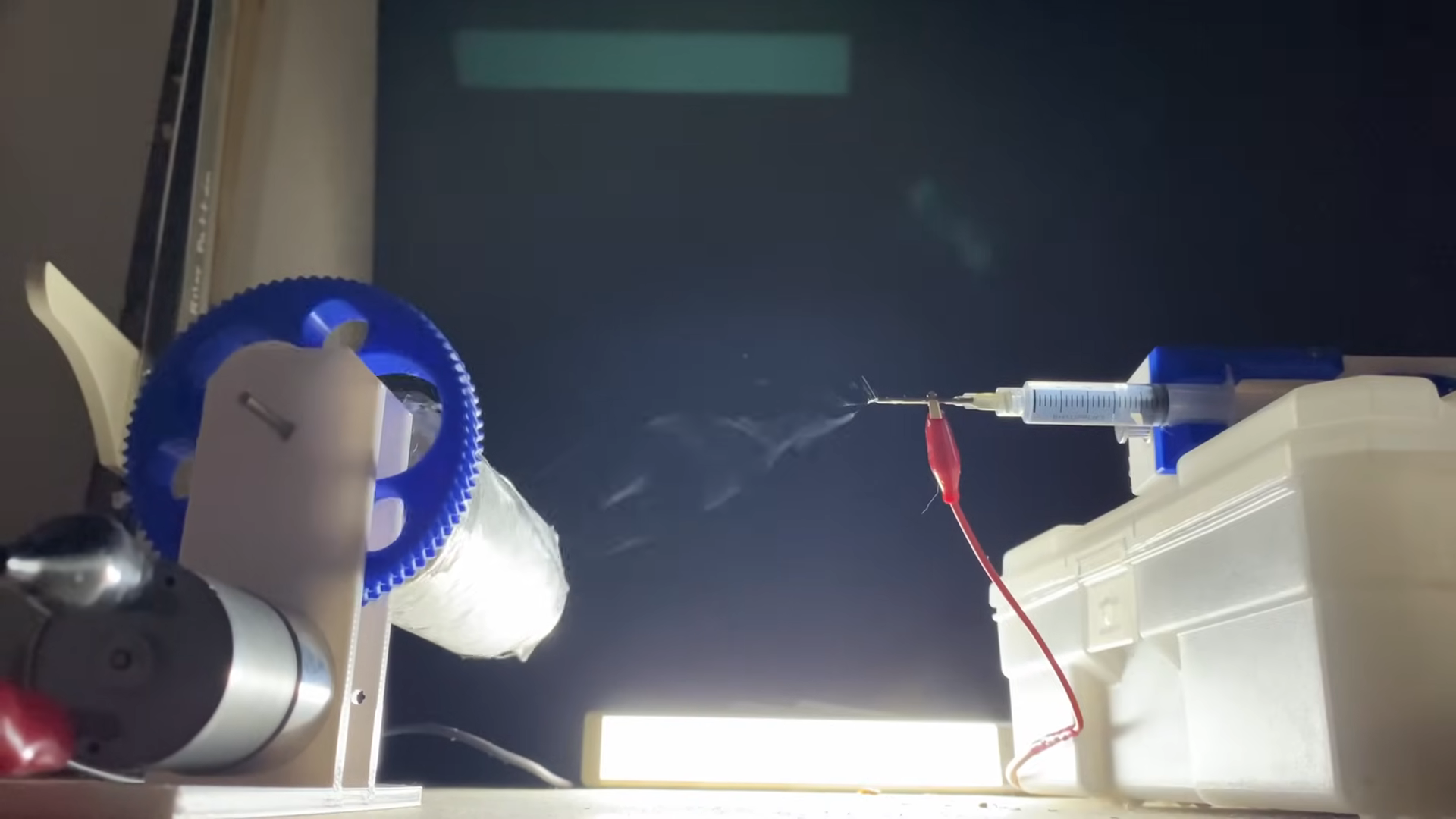
Electrospinning is a process that turns a polymer solution into extremely fine fibers using high voltage. A polymer is loaded into a syringe or reservoir. A strong electric field is applied between the liquid and a collector. When the electrical force overcomes surface tension, a thin jet shoots out, stretches, dries, and lands as a web of microfibers or nanofibers. The result can look like microscopic angel hair pasta, except it is engineered, sterile, and not recommended with marinara.
For artificial heart valves, electrospinning matters because fiber diameter, orientation, porosity, thickness, and layer structure can all be tuned. Researchers can make fibers randomly arranged for isotropic behavior, aligned for directional strength, or layered to imitate the complex structure of native valve leaflets. A healthy heart valve is not a flat flap. It is a sophisticated, multilayered tissue with collagen, elastin, proteoglycans, endothelial cells, interstitial cells, and highly organized mechanical behavior. Electrospinning gives engineers a way to imitate some of that architecture.
Why Electrospun Scaffolds Fit Heart Valve Engineering
A heart valve leaflet must be thin enough to open easily, strong enough to withstand pressure, elastic enough to snap back, and smooth enough to avoid disturbing blood flow. It also needs fatigue resistance because the human heart beats roughly 100,000 times per day. A valve that performs beautifully on Monday but gives up by Thursday is not a medical device; it is a dramatic intern.
Electrospun scaffolds offer several advantages for this challenge:
- High surface area: Thin fibers create more surface for cell attachment and protein interaction.
- Porosity: Open spaces can allow cell migration, nutrient diffusion, and tissue remodeling.
- Mechanical tunability: Fiber alignment and polymer choice can improve strength, flexibility, and anisotropic behavior.
- Biomimicry: Fiber networks can resemble the natural extracellular matrix of valve tissue.
- Drug or molecule loading: Some scaffolds can be designed to release growth factors, anti-inflammatory agents, or antithrombotic molecules.
These features make electrospun heart valve scaffolds especially interesting for regenerative medicine. Instead of simply replacing a valve with inert hardware, the scaffold may act as a temporary guide for new tissue formation.
Materials Used in Electrospun Artificial Heart Valves
The material choice determines how a scaffold behaves inside the body. It affects elasticity, degradation rate, inflammation, cell attachment, calcification risk, sterilization compatibility, and manufacturing consistency. Researchers often test both synthetic and natural polymers, as well as hybrid blends.
Synthetic Polymers
Synthetic polymers such as polycaprolactone, polyurethane, polylactic acid, polyglycolic acid, and related copolymers are popular because they are tunable and mechanically reliable. Polycaprolactone, often shortened to PCL, degrades slowly and can provide long-term structural support. Polyurethane can offer elasticity, which matters because valve leaflets bend, flex, and flutter under changing pressure. Some recent designs combine PCL and polyurethane to balance strength and flexibility.
The advantage of synthetic polymers is control. Engineers can adjust molecular weight, fiber diameter, collector speed, solvent system, and scaffold thickness to target specific mechanical properties. The disadvantage is biology. Cells may not always love synthetic surfaces at first glance. Sometimes the surface needs modification, coating, plasma treatment, or blending with natural molecules to improve cell attachment and reduce inflammatory response.
Natural Polymers
Natural materials such as collagen, gelatin, elastin, fibrin, hyaluronic acid, and decellularized extracellular matrix components can encourage cell recognition and tissue formation. Cells tend to respond well to biological cues because, frankly, cells are tiny snobs with strong opinions about their neighborhood.
The challenge is that natural polymers may be weaker, less predictable, or harder to process than synthetic materials. They may swell, degrade too quickly, or require chemical crosslinking. For a heart valve, “biocompatible but floppy” is not good enough. The scaffold must survive real blood pressure and repeated motion.
Composite Scaffolds
Many researchers now focus on composite scaffolds that combine synthetic durability with biological friendliness. A valve might use a synthetic polymer backbone for mechanical strength and a natural polymer coating for cell adhesion. Another approach is a multilayer leaflet, where each layer has different fiber orientation or material composition. This is important because native valve leaflets are not uniform. Their fibrosa, spongiosa, and ventricularis layers each contribute different mechanical and biological functions.
How Electrospun Heart Valve Scaffolds Are Made
Creating an electrospun artificial heart valve is not as simple as spraying fibers onto a heart-shaped object and calling it a day. The geometry of a valve is complex. Leaflets must meet neatly at the center, open with minimal resistance, close without leaking, and avoid wrinkling or tearing. Engineers use several strategies to get closer to that goal.
Mandrel-Based Electrospinning
A mandrel is a shaped collector that gives the scaffold its form. For tubular or valved conduits, a rotating mandrel can collect fibers in a controlled pattern. By changing rotation speed, voltage, distance, and solution flow rate, researchers can influence fiber alignment and scaffold thickness. Faster rotation may produce more aligned fibers, which can improve directional strength.
Layered Leaflet Construction
Some valve designs use electrospinning to build leaflets layer by layer. One layer may focus on circumferential strength, another on flexibility, and another on promoting endothelialization. This layered strategy attempts to imitate the native valve’s architecture, where collagen-rich regions resist stretching and elastin-rich regions help the leaflet recoil.
Combining 3D Printing and Electrospinning
One emerging approach pairs 3D printing with electrospinning. A 3D-printed frame or mold can define the valve geometry, while electrospinning creates the fibrous leaflet scaffold. This hybrid strategy can improve reproducibility and help engineers create patient-specific or size-specific designs. Think of 3D printing as building the stage and electrospinning as weaving the curtain that actually performs.
Cells, Remodeling, and the “Living Valve” Concept
The most exciting version of an electrospun artificial heart valve is not just a polymer device. It is a regenerative scaffold that becomes living tissue. There are two broad strategies: seed cells before implantation or implant an acellular scaffold that recruits cells from the body.
Pre-Seeded Tissue-Engineered Valves
In a pre-seeded design, researchers grow cells on the scaffold before implantation. These may include endothelial cells, mesenchymal stem cells, valve interstitial cells, or other cell types selected for remodeling potential. The scaffold may be conditioned in a bioreactor that mimics blood flow and pressure. This mechanical stimulation can encourage cells to produce collagen and organize tissue before the valve is implanted.
The benefit is that the valve may already have living tissue at the time of surgery. The drawback is complexity. Cell sourcing, expansion, sterility, timing, regulatory review, and cost all become more difficult. Personalized cell-based therapy is powerful, but it is not exactly a drive-through order.
Acellular Scaffolds and Endogenous Tissue Restoration
Another strategy is to implant an acellular bioresorbable scaffold. The scaffold is designed to attract the patient’s own cells after implantation. Over time, the polymer degrades while new tissue forms. This concept is often called endogenous tissue restoration. If successful, it could simplify manufacturing compared with patient-specific cell seeding while still supporting regeneration.
The challenge is control. The body’s healing response is powerful, but it can be messy. Too little cell invasion means weak remodeling. Too much inflammation can lead to fibrosis, thickening, or stenosis. The scaffold must send the right biological and mechanical signals without acting like an annoying houseguest who overstays and triggers immune drama.
Testing Electrospun Artificial Heart Valves
Before any artificial heart valve reaches patients, it must survive a long testing journey. Researchers evaluate electrospun valves through bench tests, computational models, cell studies, animal studies, and eventually clinical trials.
Mechanical Testing
Heart valve scaffolds are tested for tensile strength, bending stiffness, suture retention, burst pressure, fatigue resistance, and leaflet motion. The scaffold must be strong but not rigid. If the valve is too stiff, it may not open fully. If it is too weak, it may stretch, tear, or leak.
Hemodynamic Testing
Hemodynamics refers to how blood flows through and around the valve. A good valve should minimize pressure drop, turbulence, regurgitation, and areas of abnormal shear stress. Flow loops and pulse duplicator systems can simulate cardiac cycles to see whether the valve opens and closes properly. Computational fluid dynamics can also predict flow patterns and identify regions where clotting risk may increase.
Biocompatibility and Blood Compatibility
Because the valve touches blood constantly, hemocompatibility is essential. Researchers test whether the material activates platelets, triggers clotting, damages red blood cells, or causes inflammation. Surface chemistry matters here. A scaffold that looks lovely under a microscope but makes platelets panic is not ready for prime time.
Animal and Clinical Studies
Large-animal models, especially sheep, are commonly used in heart valve research because their cardiovascular pressures and valve sizes can provide meaningful preclinical data. These studies help evaluate tissue ingrowth, calcification, inflammation, leaflet motion, degradation, and long-term function. Only after extensive evidence can a device move toward human trials and regulatory approval.
Potential Benefits of Electrospun Artificial Heart Valves
If electrospun artificial heart valves continue to advance, they could offer several important benefits.
Better Mimicry of Native Tissue
Electrospun fibers can imitate the scale and structure of natural extracellular matrix. This may improve cell response and mechanical performance compared with solid polymer sheets or purely synthetic molded parts.
Reduced Need for Repeat Procedures
A scaffold that remodels into living tissue could potentially resist long-term degeneration better than fixed bioprosthetic tissue. This would be especially valuable for children and young adults, who may otherwise face multiple interventions over a lifetime.
Growth Potential in Pediatric Patients
One of the hardest problems in pediatric valve replacement is that children grow and artificial valves do not. A living, remodeling valve could theoretically adapt as the child grows. That would be a major breakthrough, though proving it safely will require careful long-term data.
More Personalized Designs
Because electrospinning can be combined with imaging, computer modeling, and 3D manufacturing, future valves may be tailored by size, geometry, mechanical profile, or disease type. Personalized medicine may eventually mean more than choosing the closest valve size from a shelf.
Challenges Holding the Technology Back
For all its promise, electrospinning artificial heart valves is still a difficult field. The heart is not a polite testing environment. It is wet, dynamic, high-pressure, immunologically active, and extremely unforgiving.
Balancing Strength and Degradation
A bioresorbable scaffold must stay strong long enough for new tissue to form, then degrade at the right pace. If it disappears too quickly, the valve may fail. If it lingers too long, it may interfere with remodeling or provoke inflammation. Timing is everything. This is basically the Goldilocks problem, but with polymers and cardiology.
Preventing Calcification
Calcification is a major cause of valve failure. Any new scaffold must avoid mineral buildup that stiffens the leaflets. Material chemistry, inflammation, mechanical stress, and patient age can all influence calcification risk.
Ensuring Consistent Manufacturing
Electrospinning is sensitive to humidity, solvent evaporation, voltage, flow rate, polymer concentration, and collector design. A commercial valve must be manufactured reproducibly, not “mostly similar if the lab air is feeling cooperative.” Scaling from small research samples to regulated medical devices is a serious engineering challenge.
Regulatory Complexity
Heart valves are high-risk medical devices. A regenerative valve may also involve bioresorbable materials, biologic interactions, drug-like additives, or cell-based components. That means regulatory pathways can be complex. Safety, durability, sterility, mechanical reliability, and clinical benefit must be demonstrated clearly.
Real-World Examples and Research Direction
Research groups have tested electrospun heart valve scaffolds using polymers such as PCL, polyurethane blends, PGA, PLA, and composite biomaterials. Some studies focus on pulmonary valves because right-sided heart pressures are lower than aortic pressures, making the pulmonary position a more realistic early target for regenerative scaffolds. Others explore aortic valve designs, which face higher pressure and greater mechanical demands.
Recent studies have also investigated multilayer scaffolds, aligned fibers, bioresorbable conduits, and scaffold designs that encourage endothelial coverage. The endothelial layer is important because it can help create a smoother blood-contacting surface and reduce thrombosis risk. Researchers are also paying close attention to macrophage response, because immune cells can either support constructive remodeling or drive chronic inflammation.
Another important direction is computational modeling. Engineers can simulate leaflet stress, flow behavior, fiber orientation, and tissue growth. These models help researchers predict failure points before building and testing every possible design. In a field where each experimental valve takes time and money, computer simulation is not a shortcut; it is a very useful flashlight.
Experience-Based Insights: What Working With Electrospun Valve Concepts Teaches Us
Anyone who spends time around electrospinning artificial heart valve research quickly learns that this field rewards patience. On paper, the concept sounds almost magical: spin a scaffold, implant it, and let the body rebuild a valve. In practice, every tiny detail argues back. A polymer concentration that makes beautiful fibers one week may produce beads the next. A collector that creates a lovely flat sheet may fail when asked to form a curved leaflet. A scaffold that looks perfect under scanning electron microscopy may be too stiff when tested in bending. The microscope says, “gorgeous.” The mechanical tester says, “absolutely not.”
One practical lesson is that valve engineering is not only about material strength. It is about motion. A heart valve leaflet is a moving structure, and movement changes everything. A material can have excellent tensile strength but still be wrong for a valve if it does not flex smoothly. Leaflets need to coapt, meaning they meet and seal properly when closed. Even a tiny mismatch can cause regurgitation. In valve work, a millimeter is not a small error; it is a dramatic plot twist.
Another experience-based insight is that biological success and mechanical success must happen together. A scaffold may support cell attachment beautifully but lack durability. Another scaffold may be mechanically impressive but biologically unfriendly. The best designs usually come from compromise: synthetic fibers for strength, biological cues for healing, and surface modifications to make the whole thing less suspicious to the body. The goal is not to make cells throw a party on the scaffold. The goal is to make them organize useful, durable tissue without causing inflammation, thickening, or calcification.
Researchers also learn to respect the blood-contacting surface. Blood is not passive red water. It contains platelets, proteins, immune cells, and clotting factors ready to react to roughness, chemistry, and abnormal flow. A scaffold with pores may be wonderful for tissue ingrowth, but too much roughness on the blood side can become a problem. Designing one side for cell invasion and the other for smooth blood flow is one reason multilayer electrospun scaffolds are so attractive.
Testing also teaches humility. A valve may pass static tests, then struggle under pulsatile flow. It may perform well for thousands of cycles, then reveal fatigue issues over longer testing. It may work in vitro but trigger unexpected remodeling in vivo. That is not failure; that is the scientific process doing its job, preferably before a device reaches patients.
The most exciting experience in this field is watching separate disciplines click together. Polymer chemists, biomedical engineers, cardiologists, surgeons, cell biologists, imaging experts, and regulatory specialists all have a piece of the puzzle. No single expert owns the whole answer. Electrospinning artificial heart valves is where materials science meets medicine, fluid dynamics meets cell behavior, and a tiny fibrous scaffold tries to become one of the hardest-working structures in the human body. It is ambitious, complicated, and occasionally maddeningbut the potential reward is enormous.
Conclusion: A Small Scaffold With a Big Assignment
Electrospinning artificial heart valves represents one of the most intriguing paths in cardiovascular tissue engineering. By creating micro- and nanofibrous scaffolds that mimic the body’s natural extracellular matrix, electrospinning gives researchers a way to design valves that are flexible, porous, biologically interactive, and mechanically tunable. The ultimate goal is not just another replacement part. It is a valve that can guide regeneration, remodel into living tissue, and possibly reduce the lifelong trade-offs associated with today’s mechanical and bioprosthetic options.
The technology is not ready to replace standard heart valves everywhere tomorrow morning. Major challenges remain, including long-term durability, calcification prevention, blood compatibility, degradation timing, manufacturing consistency, and regulatory validation. Still, the progress is real. From composite PCL-polyurethane scaffolds to acellular bioresorbable designs and computationally optimized leaflet structures, the field is moving from clever lab concept toward clinically meaningful innovation.
If traditional artificial valves are expertly engineered doors, electrospun regenerative valves may become something more: temporary blueprints that help the body build its own doorway. That is the promiseand the reason this tiny fiber-spinning technology has captured such big attention in heart valve research.
Note: This article is for educational and publishing purposes only. It is not medical advice, diagnosis, or treatment guidance. Patients with heart valve disease should consult a qualified cardiologist or heart valve team.