Diabetic retinopathy fundoscopy sounds like something a professor might say while pointing at a glowing eyeball diagram. In real life, it is much simpler: it is an eye exam that helps doctors look at the back of the eye, especially the retina, to check for damage caused by diabetes. And yes, it matters even when your vision seems perfectly fine.
Diabetes can quietly affect small blood vessels throughout the body, including the tiny vessels in the retina. The retina is the light-sensitive layer at the back of the eye that helps turn what you see into signals your brain can understand. When blood sugar stays high over time, those delicate vessels can weaken, leak, swell, or grow abnormally. That process is called diabetic retinopathy.
The tricky part? Diabetic retinopathy often starts without obvious symptoms. Your eyes may feel normal, your vision may look normal, and your daily life may continue as usual. Meanwhile, small retinal changes can be developing in the background like an unwanted software update. Fundoscopy helps catch those changes early, before vision loss becomes harder to prevent.
What Is Fundoscopy?
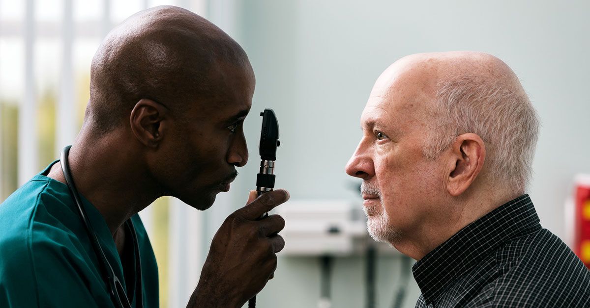
Fundoscopy, also called ophthalmoscopy or a fundus exam, is a method used to examine the fundus, which is the inner back surface of the eye. This area includes the retina, optic nerve, macula, and retinal blood vessels. During diabetic retinopathy fundoscopy, an eye care professional looks for signs of diabetes-related eye disease.
The exam may be done with a handheld ophthalmoscope, a slit-lamp microscope with special lenses, or retinal photography. In many cases, the pupils are dilated with eye drops so the doctor can see more of the retina. Think of dilation like opening the curtains wider: the view improves, but the room gets brighter than expected.
Fundoscopy is not just a quick peek. For people with diabetes, it is one of the most important tools for detecting early retinal damage, monitoring known diabetic retinopathy, and deciding whether more advanced imaging or treatment is needed.
Why Diabetic Retinopathy Fundoscopy Matters
Diabetic retinopathy is one of the leading causes of vision loss among working-age adults. The good news is that early detection and timely care can greatly reduce the risk of severe vision damage. Fundoscopy gives eye doctors a direct look at the blood vessels most likely to show early signs of trouble.
During a diabetic eye exam, the doctor may look for small bulges in blood vessels, tiny retinal hemorrhages, swelling, deposits, blocked vessels, or abnormal new blood vessel growth. These findings help determine whether retinopathy is mild, moderate, severe, or proliferative.
That staging matters because not all diabetic retinopathy needs the same response. Some people need careful monitoring and better control of blood sugar, blood pressure, and cholesterol. Others may need treatment such as eye injections, laser therapy, or surgery. Fundoscopy helps separate “keep watching closely” from “let’s act now.”
What Doctors Look for During the Exam
Microaneurysms
Microaneurysms are tiny balloon-like changes in retinal blood vessels. They are often among the earliest visible signs of diabetic retinopathy. They may be small, but they are important clues that the blood vessels are under stress.
Retinal Hemorrhages
These are small areas of bleeding in the retina. They can happen when weakened blood vessels leak. A few tiny hemorrhages may suggest early disease, while more widespread bleeding can point to more advanced retinopathy.
Hard Exudates
Hard exudates are yellowish deposits made of leaked fats and proteins. They often appear near areas of swelling. If they collect near the macula, the part of the retina responsible for sharp central vision, they can be a warning sign for diabetic macular edema.
Cotton Wool Spots
Cotton wool spots look like pale, fluffy patches on the retina. Despite the cozy name, they are not adorable. They suggest areas where the retina is not getting enough healthy blood flow.
Macular Edema
Diabetic macular edema happens when fluid builds up in or near the macula. This can blur central vision and make reading, driving, or recognizing faces more difficult. Fundoscopy may raise suspicion, but optical coherence tomography, commonly called OCT, is often used to measure retinal swelling more precisely.
New Abnormal Blood Vessels
In advanced diabetic retinopathy, the retina may grow fragile new blood vessels. This stage is called proliferative diabetic retinopathy. These new vessels can bleed or create scar tissue, increasing the risk of serious complications such as vitreous hemorrhage or retinal detachment.
Stages of Diabetic Retinopathy Seen on Fundoscopy
Mild Nonproliferative Diabetic Retinopathy
This early stage may show microaneurysms and small retinal changes. Vision may still be normal. This is exactly why regular fundoscopy matters: the eye can look fine to you while telling a different story to the doctor.
Moderate Nonproliferative Diabetic Retinopathy
At this stage, more blood vessels may show damage. Some vessels may become blocked, and the retina may begin signaling that it needs better circulation. Monitoring usually becomes more careful.
Severe Nonproliferative Diabetic Retinopathy
Severe disease means many retinal blood vessels are damaged or blocked. The retina may not get enough oxygen, which increases the risk of progression to proliferative diabetic retinopathy.
Proliferative Diabetic Retinopathy
This is an advanced stage in which abnormal new blood vessels grow on the retina or optic nerve. These vessels are fragile and can bleed. Treatment is often needed to reduce the risk of major vision loss.
What Happens During a Diabetic Retinopathy Fundoscopy Exam?
A diabetic eye exam usually begins with questions about your diabetes history, medications, blood sugar control, blood pressure, cholesterol, kidney disease, pregnancy, and any vision changes. Your eye care professional may also check your visual acuity, which is the “read the letters on the chart” part of the visit.
Next, the doctor may measure eye pressure to screen for glaucoma risk. Then come the dilating drops. These drops widen your pupils so the doctor can get a better view of the retina. They may sting briefly, and your near vision may become blurry for several hours. Bright sunlight may also feel more dramatic than a movie villain entrance, so sunglasses are helpful.
Once your pupils are dilated, the doctor examines the retina using light and magnification. The exam should not be painful, although the bright light can feel uncomfortable. Retinal photographs may be taken to document your baseline or compare changes over time.
Fundoscopy vs. Retinal Photography: Are They the Same?
They are related, but not exactly the same. Fundoscopy usually refers to the direct clinical examination of the fundus by an eye care professional. Retinal photography captures images of the retina, which may be reviewed by a trained clinician or, in some settings, supported by validated digital screening systems.
Digital retinal imaging can be useful, especially for screening programs and primary care settings. However, if images are unclear, retinopathy is detected, symptoms are present, or disease is advanced, a comprehensive dilated exam by an optometrist or ophthalmologist is still important.
Other Tests That May Be Used
Optical Coherence Tomography
OCT is an imaging test that creates cross-sectional pictures of the retina. It is especially helpful for detecting and measuring macular edema. If the macula is swollen, OCT can show where the fluid is and how thick the retina has become.
Fluorescein Angiography
Fluorescein angiography uses a special dye and camera to show blood flow in the retina. It can reveal leaking vessels, blocked areas, and abnormal new vessel growth. It is not always needed for routine screening, but it can be valuable when the doctor needs a more detailed map.
Widefield Retinal Imaging
Widefield imaging captures a larger portion of the retina than standard photos. This can help identify peripheral retinal changes that might otherwise be harder to see. It is useful in some clinics, but it does not replace clinical judgment.
How Often Should People With Diabetes Have Fundoscopy?
Screening schedules depend on diabetes type, age, pregnancy status, past exam results, and current eye findings. In general, many adults with type 2 diabetes are advised to have an eye exam at the time of diagnosis because type 2 diabetes may have been present for years before it is discovered.
People with type 1 diabetes are usually screened after several years of having diabetes, unless symptoms or other risk factors suggest earlier evaluation. Once diabetic retinopathy is found, exams are typically repeated at least annually, and more often if disease is progressing or threatening vision.
If previous exams show no retinopathy and blood sugar is well controlled, some patients may be told that screening every one to two years is reasonable. However, this decision should come from an eye care professional or diabetes care team, not from a calendar app with confidence issues.
Pregnancy changes the equation. People with type 1 or type 2 diabetes who become pregnant may need an eye exam before pregnancy or early in the first trimester, with follow-up based on findings. Gestational diabetes alone does not carry the same retinopathy screening recommendation, but anyone with vision symptoms should seek medical advice.
Symptoms That Should Not Wait
Do not wait for the next routine exam if you notice sudden vision changes. Symptoms that deserve prompt attention include new floaters, blurred vision, dark spots, sudden loss of vision, flashes of light, distorted central vision, or a curtain-like shadow in your field of view.
Diabetic retinopathy can be silent early, but advanced problems may arrive loudly. When vision changes suddenly, fast evaluation matters. The eye is not a “let’s see how it looks next month” organ when sudden symptoms appear.
Who Is at Higher Risk?
Anyone with diabetes can develop diabetic retinopathy, but risk increases with longer duration of diabetes. Poor blood sugar control, high blood pressure, high cholesterol, kidney disease, smoking, and pregnancy can also raise risk. People who already have retinopathy need closer monitoring because progression can happen even when symptoms are mild or absent.
The most practical takeaway is not to panic, but to participate. Managing diabetes is a team sport: primary care clinician, endocrinologist, eye doctor, dentist, pharmacist, family support, and yes, the person actually living with diabetes. The retina appreciates teamwork.
Can Diabetic Retinopathy Be Prevented?
Not every case can be prevented, but risk can often be reduced. Good blood sugar management is one of the most important steps. Blood pressure control and cholesterol management also help protect blood vessels. Regular exercise, not smoking, taking prescribed medications, and keeping medical appointments all support eye health.
Most importantly, do not rely on vision symptoms as your screening system. By the time diabetic retinopathy affects vision, it may already be more advanced. Fundoscopy can detect warning signs before your eyesight sends an angry email.
Treatment Options If Fundoscopy Finds Retinopathy
Treatment depends on severity. Mild diabetic retinopathy may only require better diabetes management and regular monitoring. Diabetic macular edema or proliferative diabetic retinopathy may require treatment by an ophthalmologist or retina specialist.
Common treatments include anti-VEGF injections, which help reduce abnormal vessel growth and leakage; laser treatment, which can seal leaking areas or reduce abnormal vessel growth; and vitrectomy surgery, which may be used when bleeding or scar tissue seriously affects vision. These treatments may sound intimidating, but they are routine tools in modern retina care.
How to Prepare for Your Appointment
Bring your glasses or contact lens information, a medication list, recent A1C results if available, and details about any vision changes. Ask whether your eyes will be dilated. If dilation is planned, consider bringing sunglasses and arranging a ride, especially if you are sensitive to blurred vision or bright light afterward.
Good questions to ask include: Do I have diabetic retinopathy? Is it affecting my macula? Do I need OCT or retinal photos? How often should I return? What symptoms should make me call sooner? Should my diabetes care team know about today’s findings?
Common Myths About Diabetic Retinopathy Fundoscopy
“My Vision Is Fine, So My Retina Must Be Fine.”
Not necessarily. Early diabetic retinopathy often has no symptoms. Normal vision does not always mean normal retinal blood vessels.
“A Regular Glasses Exam Is Enough.”
A glasses prescription checks how clearly you see, but a diabetic eye exam checks the health of the retina. You may need both, but they are not the same thing.
“Only People With Poor Diabetes Control Need Eye Exams.”
Better diabetes control lowers risk, but it does not erase risk. Duration of diabetes, blood pressure, cholesterol, genetics, and other health factors also matter.
“If Retinopathy Is Found, Blindness Is Inevitable.”
No. Many people with diabetic retinopathy keep useful vision for life, especially when the condition is detected early and managed properly.
Real-Life Experiences: What Patients Often Notice Before and After Fundoscopy
For many people, the hardest part of diabetic retinopathy fundoscopy is not the exam itself. It is making the appointment. Life gets busy. Work happens. School pickups happen. Insurance cards disappear into mysterious wallet caves. And because early diabetic retinopathy usually does not hurt, it is easy to delay the visit.
A common experience is surprise. Someone may walk into the clinic saying, “I see fine,” and leave with retinal photos showing early changes. That does not mean disaster is waiting around the corner. It means the exam did its job. Finding mild retinopathy early gives the patient and care team time to adjust diabetes management, schedule follow-ups, and prevent avoidable damage.
Another common experience is temporary annoyance after dilation. Reading a phone screen may become difficult for a few hours. Sunlight may feel unusually bright. A person may leave the office wearing disposable sunglasses that look like they were designed by a committee of robots. Still, most patients find the inconvenience manageable compared with the value of knowing what is happening inside the eye.
Some patients feel anxious when they hear medical terms such as “hemorrhage,” “edema,” or “proliferative.” That is understandable. Eye language can sound dramatic. A helpful approach is to ask the doctor to explain the stage in plain English: What did you see? Is my central vision area involved? Do I need treatment now, or monitoring? What can I do before the next visit?
People who have OCT imaging often describe it as quick and painless. They look into a machine, follow a target light, and the scan is completed in moments. The results can be reassuring because the doctor can show whether fluid is present near the macula. Seeing the image can make the condition feel less mysterious and more manageable.
Patients who need injections or laser treatment may feel nervous at first. That reaction is normal. However, retina specialists perform these treatments frequently, and the goal is to protect sight. Many patients say the fear before the first treatment is worse than the actual appointment. Clear explanations, numbing medicine, and knowing what to expect can make the process easier.
One practical lesson from patient experiences is to connect the eye exam with an existing routine. Some people schedule fundoscopy near their birthday, during Diabetes Awareness Month, or right after their regular primary care checkup. Others set reminders when they refill medications. The best reminder is the one you will actually follow, not the one that looks impressive in a planner and then vanishes into Monday chaos.
Family support also helps. A spouse, parent, sibling, or friend can provide transportation after dilation, help remember questions, or encourage follow-up. Diabetes management can feel lonely, but eye care does not have to be a solo mission.
The biggest experience-based takeaway is simple: do not wait until vision changes force your attention. Fundoscopy is most powerful when it catches problems early. It turns hidden information into visible evidence, and visible evidence leads to better decisions.
Conclusion
Diabetic retinopathy fundoscopy is one of the most important eye exams for people living with diabetes. It allows eye care professionals to examine the retina, detect early blood vessel changes, stage diabetic retinopathy, and decide whether monitoring, imaging, or treatment is needed. The exam may involve dilating drops, bright lights, retinal photos, OCT, or additional tests, but the purpose is straightforward: protect vision before damage becomes severe.
If you have diabetes, do not wait for blurry vision to become your reminder. Regular diabetic eye exams are a smart, practical, sight-saving habit. Your retina works hard every day. The least you can do is let someone check on it once in a while.